Broadband stimulated Raman imaging based on multi-channel lock-in detection for spectral histopathology
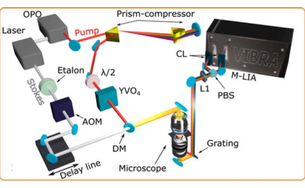
Spontaneous Raman microscopy reveals the chemical composition of a sample in a label-free and non-invasive fashion by directly measuring the vibrational spectra of molecules. However, its extremely low cross section prevents its application to fast imaging. Stimulated Raman scattering (SRS) amplifies the signal by several orders of magnitude thanks to the coherent nature of the nonlinear process, thus unlocking high-speed microscopy applications that provide analytical information to elucidate biochemical mechanisms with subcellular resolution. Nevertheless, in its standard implementation, narrowband SRS provides images at only one frequency at a time, which is not sufficient to distinguish constituents with overlapping Raman bands. Here, we report a broadband SRS microscope equipped with a home-built multichannel lock-in amplifier simultaneously measuring the SRS signal at 32 frequencies with integration time down to 44 µs, allowing for detailed, high spatial resolution mapping of spectrally congested samples. We demonstrate the capability of our microscope to differentiate the chemical constituents of heterogeneous samples by measuring the relative concentrations of different fatty acids in cultured hepatocytes at the single lipid droplet level and by differentiating tumor from peritumoral tissue in a preclinical mouse model of fibrosarcoma.